No products in the cart.

Confocal Laser Scanning Microscopes for Lab Research
Recent studies show that we can use deep learning algorithms in confocal microscopes, improving their resolution from about 240nm to 125nm. This huge increase in their already crystal-clear 3D imaging means we will likely start seeing them used for more purposes than ever.
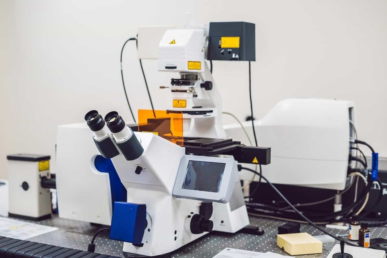
There are many types of confocal laser scanning microscopes. You should know what each one does before finding the right one for your needs.
Below, we will explore how the features of various confocal microscopes make each one a good choice for your lab research. Make better and cheaper decisions today, and make sure your device lives up to its promise of great research performance.
I. Introduction to Confocal Microscopy
Confocal microscopy has advanced significantly. It is better than older methods that used light to collect visual information about a specific area.
Instead, the process offers significantly improved resolution and clarity. It also allows for three-dimensional visualization. This is easier for institutes, especially when imaging complex samples.
The following sections detail the principles and applications distinguishing confocal microscopy from legacy techniques.
What Is Confocal Microscopy
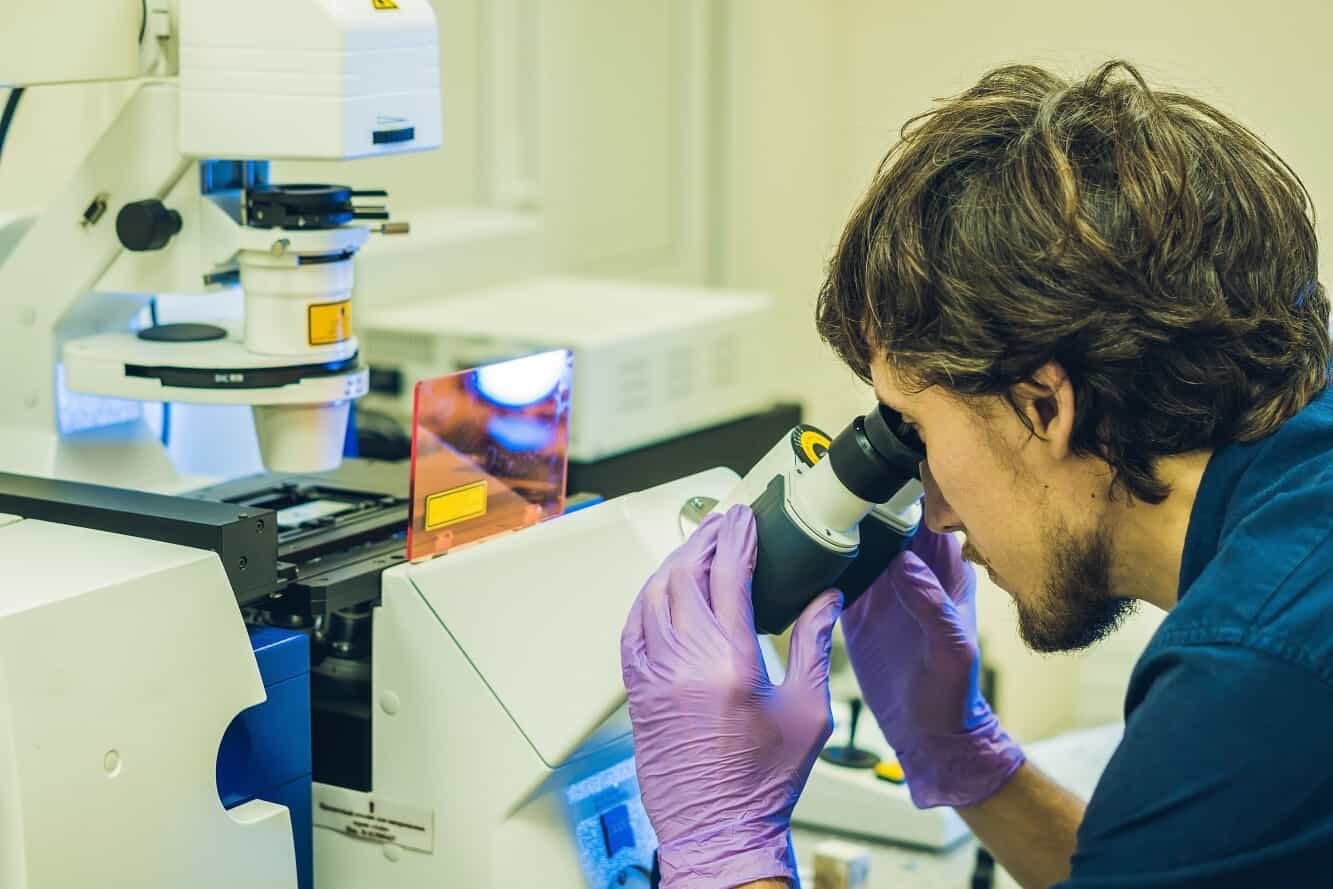
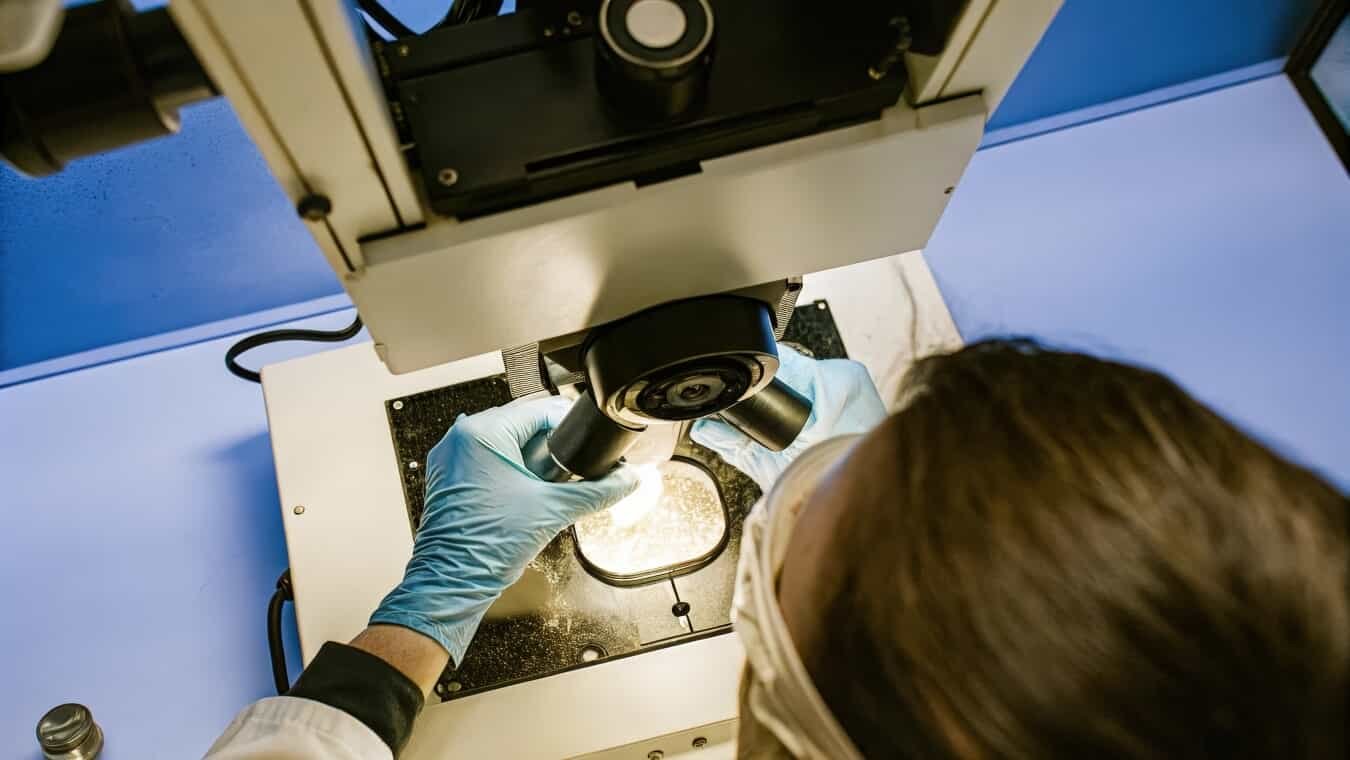
Confocal laser scanning microscopes (CLSM) use a laser beam to take images of a specific area. They use “point-by-point illumination” to light up small sections one at a time. The microscope captures as much detail as possible before moving to the next area. This allows the microscope to develop highly detailed imagery that defines abnormalities or details with greater clarity.
After scanning, a CLSM uses the information it collects to create detailed 3D images. It also makes classic slice-based visuals easier to present. Researchers can then leverage this information in a variety of fields, including:
- Cell biology
- Neuroscience
- Materials science
- Clinical diagnostics
- Semiconductor inspection
- Pharmaceutical research
- Ophthalmology
In these areas, research microscopes must show fine details in samples, and laboratory equipment should always provide high-quality results.
What Is Confocal Laser Scanning Microscopy Used For?
Researchers use CLSM technology in many areas of science, such as:
- Recording dynamic cellular processes
- Watching the movement of newly-created proteins
- Analyzing biochemical organization efforts
- Learning how cells react individually to external signals
The device can do this in real time, which helps scientists develop research that requires them to respond to how a cell reacts during their studies.
Researchers can use 3D models of complex biological structures to examine them in detail. This is important for diagnosing many diseases and assessing experimental treatments. With these details, researchers can see functions like cytoskeletal dynamics and watch how cell filaments or microtubules change during cell processes.
The CLSM can also investigate potential neurological diseases. With these research microscopes, staff can observe the brain for calcium irregularities, which helps them better understand the patient’s condition.

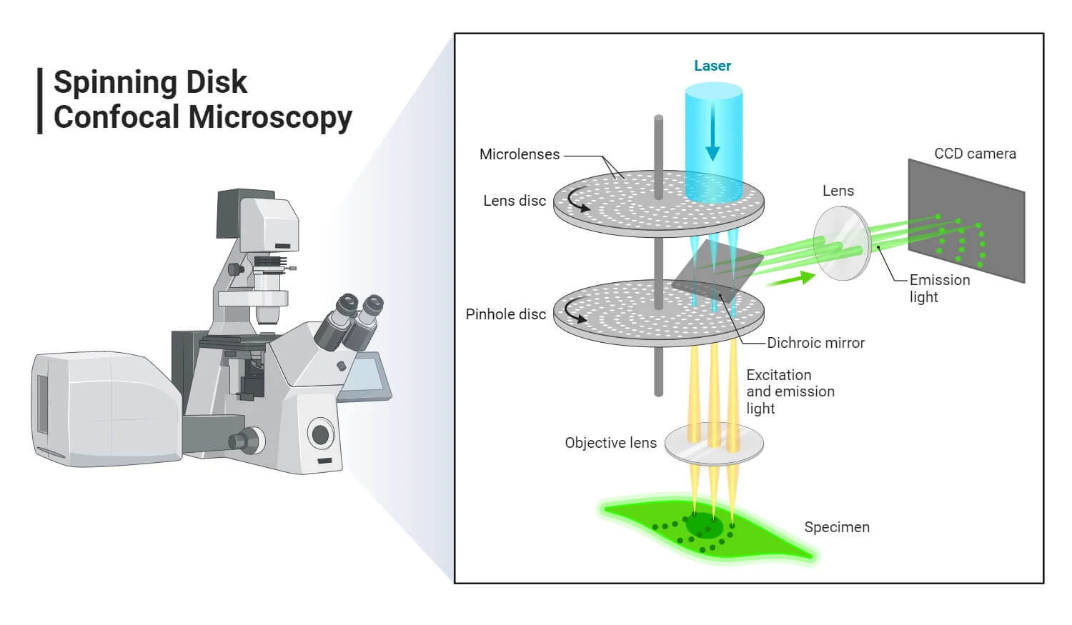
How Does Confocal Microscopy Work?
When using the CLSM, the user directs a low-power laser beam at the sample, applying it only to the area they want to study. Within the device, a pinhole positioned in the optical path blocks any light that would otherwise cause the view to lose focus. As such, it ensures that only light from the device’s plane reaches the detector.
The microscope then picks up any fluorescence emitted by material in the scanned volume. It changes light signals into clear digital images, allowing the device or connected computers to see, adjust, or analyze the results.
Specialized software can collect and combine optical images, creating a detailed 3D view of tiny structures. This is great for many research projects.
When To Use Confocal Microscopy?
Several settings provide an ideal fit for confocal microscopy. The best choice for live cell imaging, for example, is:
- Light can often kill off or harm individual cells
- Confocal microscopy offers better 3D data
- Traditional microscopes offer less depth of detail
- Thicker samples can be more challenging to analyze with other microscopes
The technology works well for imaging thick, multi-layered volumes. Precise three-dimensional reconstructions provide significant benefits. This information helps researchers and doctors understand what might happen in a specific sample.
Phototoxicity is a big problem when gathering important information about a sample. It can affect how the sample behaves. The microscope uses lasers, but it uses an incredibly small amount. It focuses the light carefully, so it is unlikely to harm the sample.

II. What Are the Advantages and Disadvantages of Confocal Microscopy?
CLSM has brought sharper and more detailed views of 3D structures to the research world. Its ability to capture high-res images in sections makes it a valuable tool for many scientific uses. However, it also comes with trade-offs.
Here, we explain the pros and cons of both areas. This will help you understand what CLSM offers and which microscopes best overcome these limitations. If you want to know more, check out our shop with a wide range of CLSM devices.
List of Advantages
Confocal laser scanning microscopes offer greater clarity than ever at a lower price. Areas where this laboratory equipment excels include:
- Boosted image contrast
- Increased resolution
- Removal of out-of-focus light
- Simultaneous multicolor imaging without needing multiple scans
- Reduced background noise and clearer data
List of Disadvantages
The main problem with CLSM is that it is a new technology. Because it is less common than other microscopes, it is often more expensive. As such, many businesses are turning to surplus providers to help them find the right microscope for their needs.
In addition to this, some of the significant challenges for CLSM users include:
- Regular maintenance and calibration to preserve image quality
- Longer image acquisition time may hinder more extensive studies
- High-speed cellular processes may not be captured
- Device complexity may demand more extensive training
- Higher floor for technical expertise in the device’s use
- The space and environmental needs of a CLSM device
Some organizations with limited funds may find these challenges difficult to overcome. However, as technology becomes more available, Manzo Eye Care will be better able to supply it. This should give all locations access to these devices.
III. A Comparison of Confocal Laser Scanning Microscopes
As a researcher, you should take the time to choose a system best suited for the types of specimens you wish to investigate. No matter your organization, you can always make mistakes when buying equipment.
Such errors can cause significant challenges after acquiring a device as complex as a CLSM. Take your time to understand each option. See how it can help you and how it compares to others on the market.
When purchasing a confocal microscope, you must consider the potential for future expansion. So, check if you can pick up a modular or scalable CLSM. Then, please work with the designer or manufacturer to improve its parts over time. This will keep your investment relevant as new technology comes out.
1. ZEISS LSM 980 with Airyscan 2 ($59,999.00)
The Zeiss LSM 980 is a powerful device. Its Airyscan 2 detector can create detailed images faster than most traditional confocal microscopes. The technology also minimizes the amount of wasted light it produces, making images clearer and sharper.
The creators claim it can detect fluorescent markers from 380 nm to 900 nm, which allows one to see many parts of a sample simultaneously.
Zeiss’ software can automate the imaging process. It uses AI sampling to make experiments more efficient.
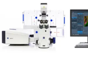
The manufacturers take pride in their ability to find exceptionally weak signals. This helps them study smaller details in cells and gives them better chances for high-quality results. The device works well for both live cells and fixed samples, too.
If this is your first time using CLSM, remember that your staff will need to learn these advanced features a lot. However, when they are up to speed, they have an even greater capability to achieve better results.
The technology within the ZEISS LSM 980 with Airyscan 2 is not compact. The large and bulky device demands a lot of dedicated lab space and regular maintenance. You should expect to fix issues with it more often than with a more established microscope. The older model has had its bugs and problems worked out.
2. ZEISS LSM 900 with Airyscan 2 ($39,500.00)
As an older piece of technology than the 980, the LSM 900 with Airyscan 2 is a much more compact and efficient version of its younger cousin. The Airyscan 2 technology is powerful, and you can still expect high-quality images from the device.
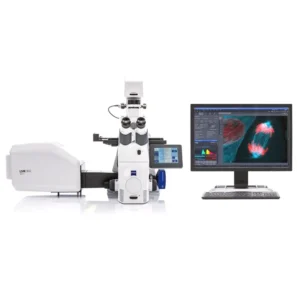
The 900 is more affordable and easier to use than later models. This simplifies investing and reduces the time between buying and using it. Onboarding new staff will also become easier because trainers will need less time for training.
In general, the 900 is a reliable device for everyday use. However, it is also important to remember that this means that it is not quite as powerful as the 980. It cannot provide as much flexibility or adjustability. The imaging is also a bit slower, which may not work for some research.
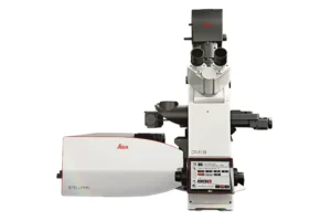
3. Leica Mica Microhub ($39,900.00)
The Leica Mica Microhub is an all-in-one imaging device that combines widefield and confocal capabilities in one system. If you don’t need the exact details of the above options, you can work faster and more simply to get the desired results.
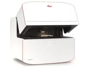
Be aware, however, that the Leica Mica Microhub comes in four different versions. They offer different capabilities:
- Live cell imaging support
- Confocal illumination
- Environmental control
- Immersion dispersion
- Imaging modes
Ensure you have all the information on each before you finalize a purchase.
Because of these differences, the Microhub does not have as much flexibility as many other systems. Its capabilities may be limited, meaning you may need to upgrade sooner.
4. Leica STELLARIS Cryo Confocal Microscope ($35,000.00)
The Leica STELLARIS Cryo Confocal Microscope makers designed it specifically for cryogenic applications. The intent is to allow it to preserve vitrified samples and maintain constant cryogenic conditions during imaging.
The device uses LAS X Coral Cryo software, which helps achieve precise 3D targeting at the nanometer scale. It works well even with temperature changes and other environmental factors.
The Cryo Confocal Microscope has ice-checking modes, which help you monitor ice thickness while you test.
The maintenance and calibration of a heating device is more demanding and may cost more. So, ensure you are prepared to respond to issues this may cause.
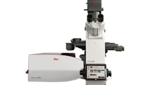
5. Leica STELLARIS 8 Confocal Microscope ($15,900.00)
The STELLARIS 8 is Leica’s newest confocal device for research. It uses an extended White Light Laser (WLL) for better image capture and detectors that work efficiently in the spectral range of 410-850 nm.
Leica knows that using CLSM machines can be challenging for most researchers. To simplify experiments, they have created an easy-to-use interface. This benefit can help ensure more reliable images while speeding up research productivity.
However, the main issues are this device’s advanced detection and laser capabilities. These features can be expensive for smaller laboratories, and their ongoing maintenance can be more intensive. So, you may need to increase your budget to compensate.
Conclusion
Confocal microscopes can transform lab research because of their high resolution and advanced imaging capabilities. Investing in advanced systems like those from Zeiss or Leica can be beneficial, but they also come with increased costs. As such, you may want to look into refurbished models.
We provide CLSM options that perform well. Our microscopes can help you save money while still ensuring quality. We check everything we buy to ensure it meets the most rigorous requirements. So, check out your options at Manzo Eye Care to find a reliable, certified, and technically powerful microscope today.
[simple-author-box]